


CT fo sinusitis
2 مشترك
صفحة 1 من اصل 1

 CT fo sinusitis
 من طرف Shamsology السبت أغسطس 28, 2010 11:21 pm


The
following is designed to enable you to develop a basic understanding of
sinus anatomy as well as CT scans, both normal and abnormal. We will
review several CT scans, but start with a drawing to start to orient
you.
There are four sets of sinuses: maxillary, ethmoid, frontal and sphenoid
sinuses. We will examine most of them in the following series of
drawings and CT scans. The initial concepts are a little difficult to
understand, but will become clearer when we get to the CT scans.

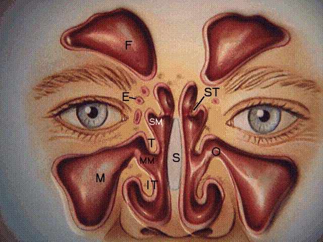
LEGEND:
F - Frontal sinuses, E - Ethmoid sinuses, M - Maxillary sinuses, O -
Maxillary sinus ostium, SS - Sphenoid sinus ST- Superior turbinate, T -
Middle turbinate, IT- Inferior turbinate, SM- Superior meatus, MM-
Middle meatus, SR - Sphenoethmoidal recess, S- Septum, ET - Eustachian
tube orifice, A - Adenoids . Courtesy of Astra Pharmaceuticals
In the first graphic representation, the three overlapping flaps of
tissue, called turbinates (inferior - IT, middle - T, and superior - ST )
protect the openings of the sinuses, and allow humidification,
filtration and warming of air. The frontal (F) sinus is seen in this
view, but is not usually involved to any great extent in sinusitis. The
sphenoid sinus (SS) is also seen in this view, and is sometimes involved
in sinusitis. The sphenoid sinus drains into the sphenoethmoidal recess
(SR)
In the second graphic representation, the maxillary sinuses (M) drain
through the maxillary sinus ostia (O) into the middle meatus (MM). It
should be noted that in this graphic diagram, the opening at O appears
to be extremely large. In actuality, it is the size of a pin head and
actually follows a rather circuitous route as you will see on the CT
scans which follow. The ethmoid sinuses (E) drain into both the middle
meatus as well as into the superior meatus (SM).
The middle meatus (MM) is bounded by the middle turbinate (T) and the
inferior turbinate (IT). (There is also a superior turbinate (ST), but
that is relatively unimportant.)
Another important structure is the "ostiomeatal unit" which is
the outflow tract from the sinuses and includes the ostium of each sinus
as well as the meati. When blocked, the ostiomeatal unit can cause
obstruction of the sinuses, analogous to putting a plug in a bathtub.
The frontal sinuses (F) are occasionally important, but will not be
dealt with to any great extent in this discussion. The septum (S)
creates a barrier between the two sides of the nose. If it is deviated
to a great enough extent, an obstruction can occur. Occasionally, there
may be a perforation (hole) in the septum, which can cause problems with
the architectural support of the nose.
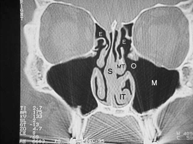
The first CT scan is relatively normal:
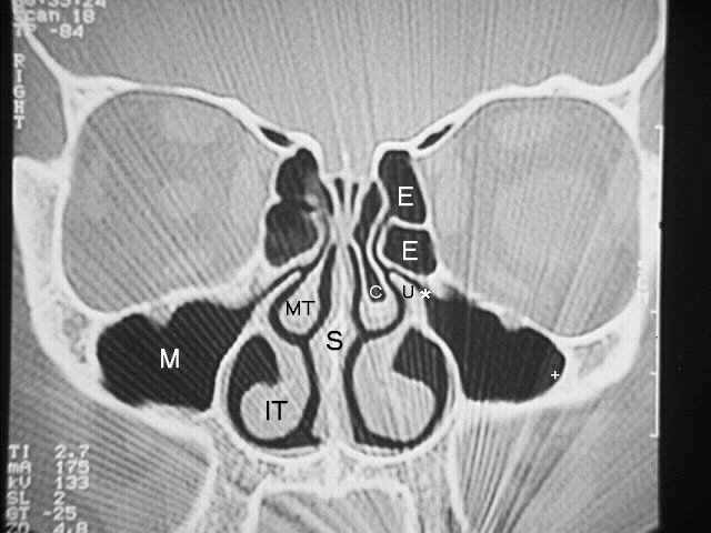
LEGEND:
+ - border of maxillary sinus, * - maxillary sinus ostium, U - uncinate
process, E - ethmoid sinuses, IT- inferior turbinate, MT- middle
turbinate, S - septum, C - concha bullosa.
Note that the CT scan is a computerized X-Ray taken in the same way as
the first diagram is drawn, as if you were able to look head on into the
sinuses. Note that the patient's right side is on your left as
indicated by the "right" mark in the upper left hand corner.
On the CT scan, bone appears white, air appears black, and soft tissue,
fluid, or muscle is varying shades of gray. Of note in the bottom
portion of the scan is a ray pattern emanating from the teeth. This is
as a result of poor penetration of the x-rays through the metal in the
teeth.
When we evaluate the sinuses for sinusitis, we look for thickening of
the lining of the sinus. Note on the patient's left side (your right) at
the + sign, there is a very sharp distinct border between the black air
in the maxillary sinus and the white bone. As you will see later,
sinusitis is manifested by grayish thickening of the lining of the
sinus.
The asterisk (*) is at the point where drainage occurs from the maxillary sinus into the nose through part of the ostiomeatal unit.
The maxillary sinus ostia is bounded below by the uncinate process (U)
and above by the lower bony portion of the ethmoid sinuses (E). A
narrowing in this area obviously can be very critical. The ethmoid
sinuses, as can be seen, are much smaller than the maxillary sinuses.
On the right side (your left), one can see the middle turbinate (MT) as
well as inferior turbinate (IT). There is a slight deviation of the
septum (S) to the right side (your left), but in this case it is
unlikely that it is causing any obstruction. Of note is that there is
air contained in the middle turbinate on the left (C-short for concha
bullosa). This represents a normal anatomical variant in which the
ethmoid sinuses have pushed down into the middle turbinate. In this
case, it does not appear to have caused a problem, but often it will
cause a significant enlargement of the middle turbinate and consequently
an obstruction on one side of the nose.
The next CT scan is from a patient with significant sinus disease.
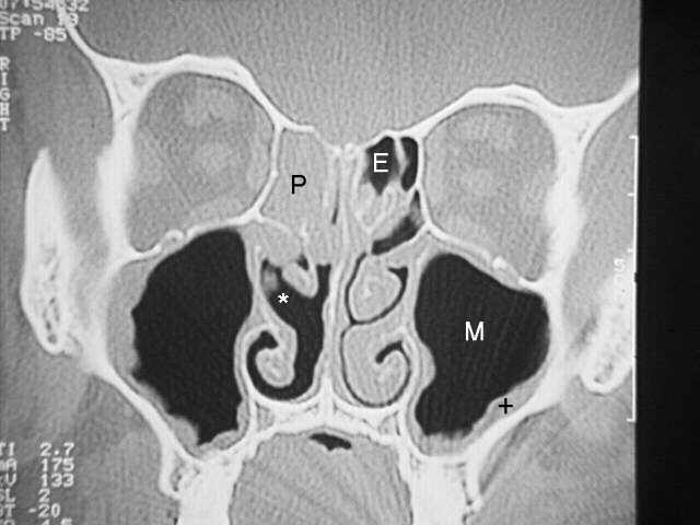
LEGEND:
M - maxillary sinus, + - thickening of the maxillary sinus, E - ethmoid
sinuses, P - polyp, O - maxillary sinus ostium, * - middle meatus.
Attention should first be directed to the + sign on the right side.
Compared to the previous scan, there is a significant amount of grayish
thickening between the white bone and the black sinuses. Any thickening
over 3 mm is definitely abnormal. Note that this thickening involves
almost the entire maxillary sinus on both sides, but more so on the
right side.
Compare the area on the right side where the maxillary sinus ostium (O)
was observed on the previous CT scan. There is no opening now, only the
gray tissue completely blocking the ostium. Not surprisingly, this
patient had a great deal of pain as a result of that blockage. Although
there is more thickening of the sinus lining on the right, there is more
room to breathe through the nose on the right side (*) than on the left
side. This is largely as a result of the deformity of the middle
turbinate, located just above the asterisk (compare to the opposite
side). This may have contributed to the sinus disease in this case,
causing obstruction of the ostiomeatal unit. Not surprisingly the
ethmoid sinuses were involved as well. The ethmoid sinuses are either
filled with polyps (P) or the lining is thickened. There is very little
air left in the ethmoid sinuses (E).
This X-Ray is from another patient with fairly severe sinus disease:
LEGEND:
MT- middle turbinate, IT- inferior turbinate, P - polyp or cyst, E -
ethmoid sinuses, O - maxillary sinus ostium, * - frontal sinus.
As compared to the previous x-ray, this patient has larger polyps or
cysts (P) (it is often difficult to tell the difference on CT scan) on
the right side, with significant obstruction of the ostium. On the left
side the ostium (O) cannot be clearly seen, but you get the idea of
where it's supposed to be. The ethmoid sinuses (E) on the right side are
completely obstructed, being filled with either polyps, cysts or
thickening of the sinuses. On the left side, there is some thickening,
but as you can see, for the most part the ethmoid sinuses are fairly
clear. The asterisk on the left side represents the point at which the
ethmoid sinuses merge into the frontal sinuses. Note the difference in
size between the middle turbinate (MT) on the right and left side, and
also the inferior turbinates (IT) on each side.
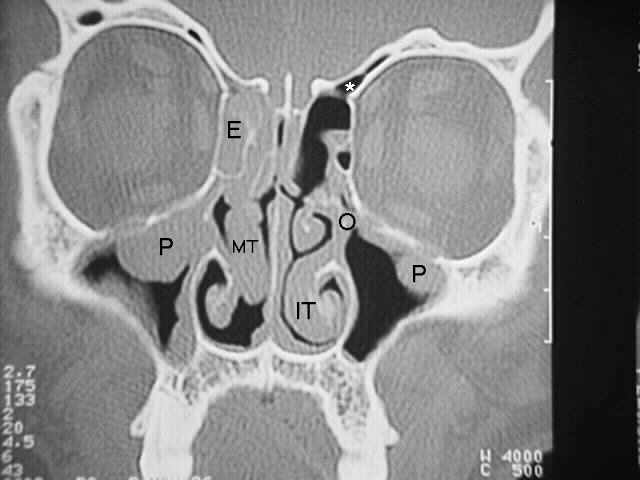
This CT scan was done after surgery was performed on the patient
whose CT scan you just reviewed.
LEGEND:
E - Ethmoid sinuses, O - maxillary sinus ostium, M - maxillary sinuses,
S - septum, MT - middle turbinate, IT - inferior turbinate.
That's pretty impressive isn't it ?, even if you're a lay-person. As you
can see, the polyps that were previously in the maxillary sinus (M) are
now gone and the opening at the maxillary sinus ostia (O) is wide open,
having had the uncinate process removed. The ethmoid sinuses (E) on
both sides have been cleaned out. The surgery which was performed did
not involve extensive removal of the lining of the sinuses. Just opening
them up and allowing them to "breathe" is often enough to prevent
severe disease. Of note, however, is the fact that this patient still
doesn't have a normal nasal airway. (Compare to the first CT scan.
Note the size of the black area next to the middle turbinate (MT) and
inferior turbinate (IT).) In addition to having sinus problems, this
patient also has allergy problems which had to be treated in order to
prevent future nasal problems and sinus disease. We hope that the short course on X-Rays of the sinuses has been helpful in understand a little more about sinusitis

Shamsology- مـشــرف عــام

- عدد المشاركات : 1191
تاريخ التسجيل : 16/07/2010
المود :


طب عين شمس- الإدارة

- عدد المشاركات : 1480
تاريخ التسجيل : 24/08/2010
المود :
صفحة 1 من اصل 1
صلاحيات هذا المنتدى:
لاتستطيع الرد على المواضيع في هذا المنتدى